Enucleation and Evisceration

Engraving diagram of the human eye and surrounding structures, numbered 1 through 7 for anatomical reference.
Illustration of a human eye cross-section showing ocular muscles, optic nerves, and surrounding bone structure. Emphasizes anatomy and function.

What is Enucleation?

Enucleation is the removal of the whole eye, including the contents (lens, uvea, etc) as well as the lining (sclera, cornea). The muscles are detached from the eye before the eye is removed.

Results

Close-up of a child's eyes with brown irises and prominent eyelashes. The right eye is turned outward, indicative of strabismus. The expression is neutral.

Enucleation

Cross-section diagram of the eye showing the optic nerve, extraocular muscles, Tenon’s capsule, and eyelids in labeled detail.
Illustration of an eye socket with a labeled implant. The implant is a textured gray sphere surrounded by yellow tissue and red muscle strands.
Cross-section illustration of an eye orbit, showing a gray prosthesis sphere surrounded by yellow tissue. Labeled "Prosthesis" with an arrow pointing to it.
Cross-section illustration of the eye shows extraocular muscles, eyelids, optic nerve, and Tenon's capsule with labels. Bright, detailed, educational.
Diagram illustrating the placement of a spherical ocular implant within the orbital socket next to a stylized eye structure.
Diagram of the human skull from above showing eye sockets and optic nerves. Labels indicate measurements and anatomical structures like the optic chiasm.

It is amazing! I love...

63 Yrs Old with Fat Droopy Eyes - Salt Lake City, UT

Well, the last time I had a picture taken I could hardly see my eyes because of the weight of heavy eyelid. Then I paid attention to how I was actually using my eyes and I really noticed when I was looking at anything especially the computer I was straining my forehead to see better. Since I have had it done I no longer have to lift the forehead and tilt my head to see.

D. Rock

Evisceration

Evisceration is the removal of the contents of the eye whilst leaving the scleral lining with the muscle attached intact:

Illustration of an eye with surgical retractor exposing a blue iris and red, vein-filled sclera. The image conveys a clinical, medical tone.
Alt text: "Illustration of cataract surgery, showing an eye held open by speculums with a scalpel cutting into the lens, highlighting the surgical process."
the corneal button is removed
Illustration showing a cross-section of an inflamed eyelid with a chalazion. A scalpel approaches the swollen area, highlighting surgical treatment.
removal of the cornea
Illustration of eye surgery shows the eye held open by a speculum, with surgical forceps gently grasping the edge of the cornea for precision operation.
Incision of the conjunctiva and Tenon’s lining
Illustration of eye surgery shows the eye held open by a speculum, with surgical forceps gently grasping the edge of the cornea for precision operation.
the Tenon’s and conjunctiva are undermined up to the equator to allow the tissues to be mobilized forwards.
An illustration of an eye surgery in close-up, showing eyelid retractors holding the eye open, with surgical tools operating on the cornea.
the corneal button is removed, followed by dissection within the sclera to remove the intraocular contents which include the lens, the choroid, the retina, etc.
Illustration showing a cross-section of an inflamed eyelid with a chalazion. A scalpel approaches the swollen area, highlighting surgical treatment.
Posterior dissection behind the sclera.
Illustration of eye surgery showing tools holding open an eye, with a lens being adjusted. It conveys a clinical, precise medical procedure.
dissection posteriorly to remove the intraocular contents
Illustration of a hernia surgery showing a red tissue protruding through an abdominal opening. Surgical tools hold the tissue, surrounded by yellow fat and muscle layers.
Evisceration being completed with removal of the attachments at the optic nerve head
A diagram of an eye surgery shows the eyelids held open with retractors. A surgical tool touches the eye, depicting a medical procedure.
the sclera is “scrubbed” to ensure that no uveal tissue is left behind.
Cross-section illustration of an ear canal with a cotton swab inserted, emphasizing ear anatomy. The swab touches the ear drum, conveying caution.
Cleaning the sclera, followed by relaxing incisions of the sclera to allow the placement of the correctly-sized implant
Medical illustration of an eye surgery with retractors holding eyelids open and surgical instruments placing a disc on the eye, indicating precision.
Placement of the implant which may be made of hydroxyapatite or polyethylene
Illustration of tweezers removing a silver circular object from an ear canal cross-section, highlighting anatomical features in vivid colors.
The positioning of the implant is helped by performing relaxing incisions in the posterior sclera
A detailed medical illustration showing a surgical procedure with an open incision being stitched. Surgical tools and retractors are visible, indicating precision.
Layered closure of the Tenon’s fascia and the conjunctiva
Cross-section illustration of a human eye with a cataract. The image shows ocular anatomy, highlighting the lens with cloudy discoloration.
Closure is performed without tension
A close-up of a closed eye with four small green plastic devices attached around it, possibly for medical or educational purposes, set against skin tone.
Temporary tarsorrhaphy sutures are placed
A close-up of a closed human eye showing redness around the eyelid, suggesting irritation. A ventilated protective eye shield with holes is visible beside it.
a conformer is placed in the fornices
Close-up of a person's face with a medical eye patch over the right eye, revealing only the left blue eye. The tone is clinical and focused.
a patch is placed on the eye

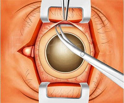
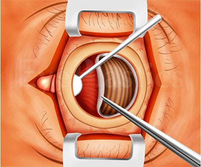
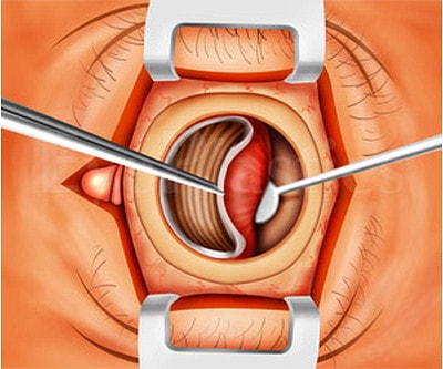
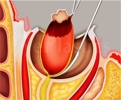
What types of implants are placed in the orbit?

Historically, implants have been made of many materials: magnets, gold, silver, glass, silicone, cartilage, bone, fat, cork, titanium mesh, acrylics, wool, rubber, catgut, peat, agar, polyethylene, hydroxyapatite.

The modern options include the following:

MEDPOR®

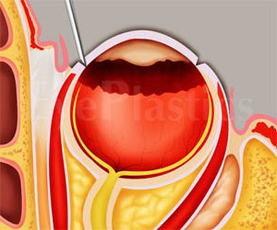
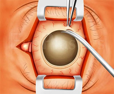
MEDPOR® is comprised of a lightweight, porous form of high-density polyethylene, a material with a long history of medical applications. Its unique, highly porous texture allows vessels to incorporate into the enhancement shape, integrating MEDPOR® 
into a patient’s tissues. The shape and size can be customized by your surgeon to fit your individual needs. MEDPOR® eliminates the need for grafts or silicone implants.

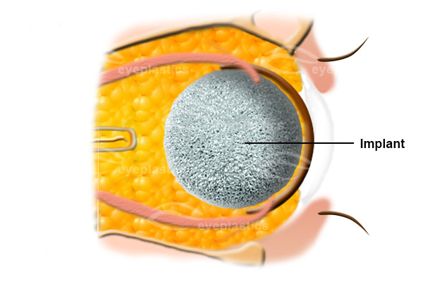
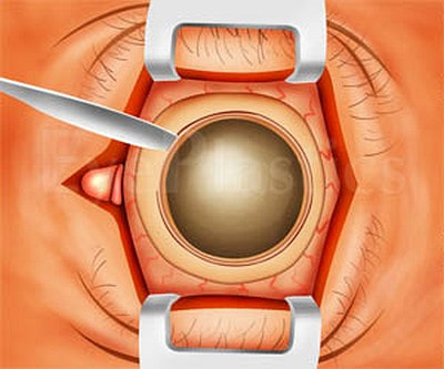
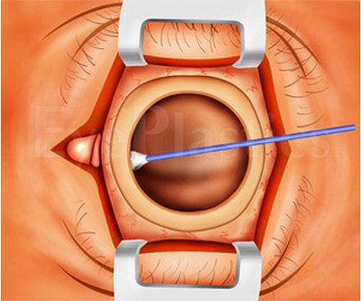
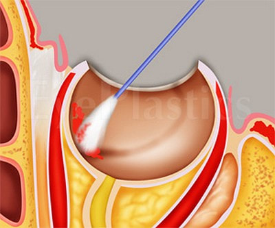
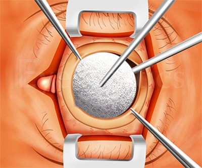
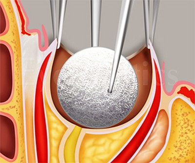
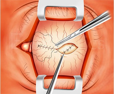
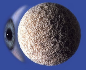
MEDPOR® Orbital Spheres

A white sphere in the center of a dark, maroon background, creating a stark contrast. The sphere casts a subtle shadow, adding depth and simplicity.

We select from sphere implant diameters of 18 mm to 22 mm.

​A resterilizable sizer set is available for selecting the appropriate implant diameter at the time of surgery.
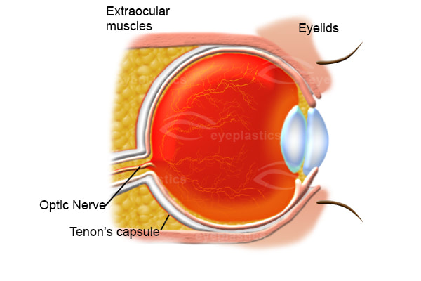
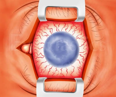
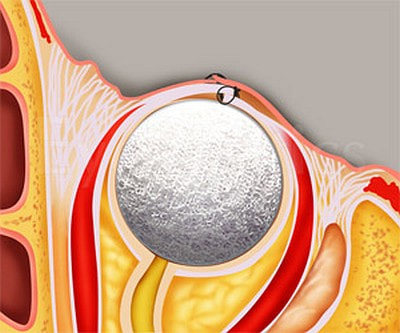
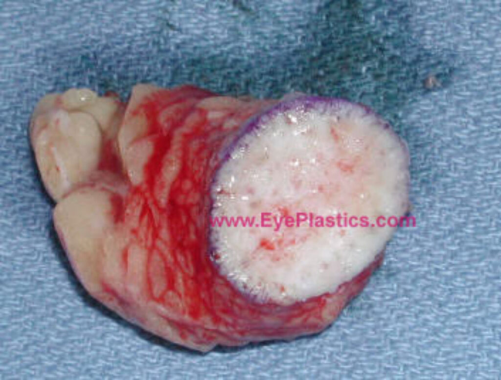
Hydroxyapatite:

Composite image of an eyeball partially covered by a textured sphere resembling a cantaloupe, against a solid blue background. Surreal and intriguing vibe.
  • The Bio-eye hydroxyapatite (HA) ocular implant is a spherical (ball-shaped) implant composed of natural coralline HA.
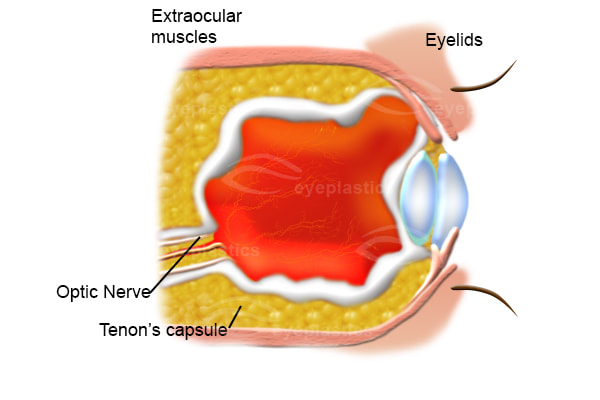
  • It is used to replace the volume of the orbit when the eye is surgically removed, or as a replacement implant in patients with a poorly functioning, pre-existing implant.
  • Historically, the use of nonporous synthetic ocular implants has led to complications such as exposure, extrusion, migration, infection, poor motility, and poor cosmesis

Dermis-fat graft

  1. is composed of subcutaneous fat and overlying dermis.
  2. Its advantages include the fact that it is an autologous graft and, thus, lacks concerns for bio-compatibility in disease transmission. However, there is a certain degree of fat atrophy which might possibly occur leading to somewhat unpredictive and result of volume.
  3. Dermis fat graft might possibly be used as a primary procedure following primary reconstruction for orbital exenteration, as a low cost means to provide an orbital implant and in instances in which enucleation is performed early in childhood since the fat might possibly grow and provide stimulus for orbital growth.
  4. Dermis fat graft can also be used in numerous secondary procedures such as a replacement of an extruded orbital implant and correction of deep superior sulcus deformity.
  5.  The surgeon might possibly choose one of several donor sites including the lower abdominal quadrant and the outer ​
  6. quadrant of the buttock.
  7. Typically a graft is measured approximately 25 mm in diameter. The epidermis might possibly be abraded with the derma-brader or with a #15 blade.
  8. ​The graft is harvested with an attempt to minimize mechanical manipulation of the graft and to quickly insert the graft into the previously prepared recipient bed.
  9. Often the dermis has slight surgical modifications in order to create a dome shape to it which often provides better motility for the prosthetic implant.
A specimen of an excised tumor with a white circular end and red, veined sides, placed on a textured blue fabric background. The image is clinical.
Close-up of a stitched surgical incision on skin, surrounded by faint markings from surgical planning. The area appears clean and healing.
A white sphere in the center of a dark, maroon background, creating a stark contrast. The sphere casts a subtle shadow, adding depth and simplicity.
Composite image of an eyeball partially covered by a textured sphere resembling a cantaloupe, against a solid blue background. Surreal and intriguing vibe.

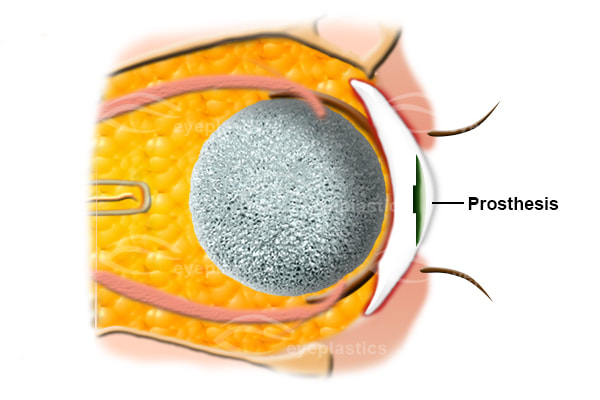
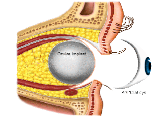
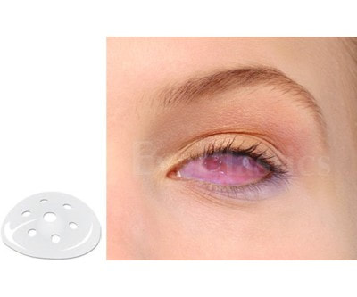
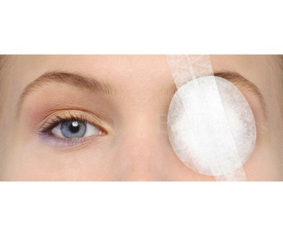
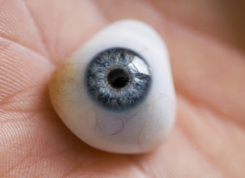
What is the prosthetic eye made of?

Close-up of a hand holding a realistic prosthetic eyeball. The eye is blue with a detailed iris and pupil, creating an intriguing and lifelike appearance.
Prosthetic eyes used to be made of glass. Because of the incidence of breakage, most modern prosthetic eyes are made of polymethylmethacrylate. The prostheses are polished every six months or so by the prosthetician.

Visit our YouTube channel for expert tips and guidance

Schedule a Consultation Today!