



While under-eye fillers are generally considered safe, they do still have a profile of potential side effects. These potential complications are tied to the delicate anatomy of the under-eye area, which requires in-depth understanding to foster safe, complication-free treatments. Below, we cover the most common complications of under-eye fillers and how they can be avoided.
Common Complications of Under-Eye Fillers
Nonsurgical procedures, such as nonsurgical under-eye bag removal, have a high safety profile for appropriate candidates, with an emphasis on “appropriate candidates.” In-depth consultation with a qualified, experienced medical professional should always be the first step in the process. Following the consultation process and working with a skilled physician are the two best ways to reduce the risk of each of the complications below.
1. Swelling and Bruising
While more of a side effect than a complication, swelling and bruising are common after filler injections. The under-eye area has thinner skin and more vascularity, which can make bruising more noticeable.
Most patients should have no more than 3–7 days of bruising, though some minor discoloration can persist past this point. If bruising persists beyond 2 weeks, we recommend an evaluation.
How to Minimize the Risk:
- Avoid blood thinners 1 week before treatment
- Pause certain supplements 1 week before treatment (e.g., vitamin E, fish oil, garlic, etc.)
- Avoid alcohol 48 hours before treatment
- Select a skilled injector
2. Lumps, Bumps, and Uneven Texture
The most common cause of uneven texture and bumps is when under-eye filler is not properly administered, such as:
- Filler being too superficially placed
- Uneven distribution
- Excess product administered
- Temporary swelling around the filler
How to Minimize the Risk:
Avoiding bumps and texture issues largely depends on the choices your injector makes. They must properly decide on:
- Product type
- Injection technique
- Aftercare instructions
3. The Tyndall Effect or Bluish Discoloration
The Tyndall effect describes how light scatters when hitting particles in a transparent medium. With under-eye fillers, it can occur when hyaluronic acid filler is placed too close to the skin surface. The area can develop a blue or gray tint due to light scattering off the clear filler gel after passing through the skin.
How to Minimize the Risk:
Your injector must apply careful techniques such as:
- Proper filler placement depth
- Microdroplets rather than large pools of filler
- Use of cannula over needles
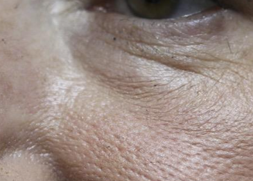
A bluish hue with swelling in front of the septum. Filler is too superficial, leading to the “Tyndall” effect
4. Overfilling and Under-Eye Puffiness
Too much filler can lead to a swollen or unnatural look to the under-eye area. What first might be mistaken for persistent swelling could actually just be too much filler. Additionally, some HA fillers can attract water. Results may look natural at first, but without considering this factor, an injector may have actually overfilled the area.
How to Minimize the Risk:
- Conservative, strategic filling
- The surgeon should be willing to under-fill and then slowly increase the fill as needed
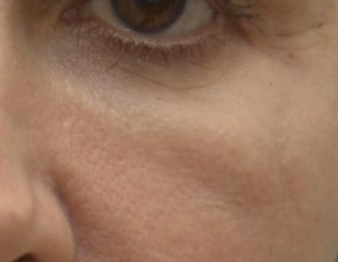
Filler causing malar edema with an oblique bulge below the eyelid. This indicates injection of too much filler, too superficial filling and filling in the wrong plane.
5. Filler Migration
If filler spreads to the area beyond the intended area, it can lead to issues like puffiness, fullness, or contour irregularities.
How to Minimize the Risk:
- Proper evaluation of filler placement
- Accurate assessment of the patient’s underlying anatomy
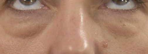
“Filler Bags”: indicate too much filler injected in the wrong plane causing the lower eyelid puffiness to look worse
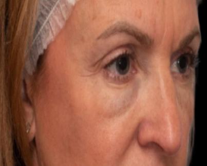
Another example of a “Filler Bag”. This causes the lower eyelids to look more puffy and the degree of puffiness will vary as fluid accummulation continues to vary. There is a malar groove and a zygomatic groove. This would require dissolving of the filler, removal of any residual filler and a “Hammock Lift” with correction of the lower eyelid laxity, skin excess, correction of the malar droop, and nano fat grafts to fill the malar and zygomatic grooves
6. Infection or Inflammatory Reactions
With noninvasive treatments, infection or delayed inflammatory reaction is a very rare occurrence (rates as low as 0.02%). Still, patients should be aware of the risk and look out for warning signs such as:
- Increasing redness
- Warmth
- Pain
- Significant swelling
- Drainage or fever
How to Minimize the Risk:
- Experienced injector techniques
- Proper aftercare for the injection sites
7. Vascular Occlusion
Even more rarely, vascular occlusion can occur (rate of < 0.1%), which entails filler entering or compressing a blood vessel. These complications are very rare with lower eyelid injections. Warning signs include
- Severe or unusual pain
- Skin blanching
- Mottled discoloration
- Visual symptoms
Of note, any sudden vision changes after filler injection require immediate emergency medical attention.
How to Minimize the Risk:
- Injector with comprehensive knowledge of facial anatomy
- Prompt recognition of complications
How Under-Eye Filler Complications Are Corrected
Depending on the complication, the corrective approach can differ. Minor issues like swelling are generally self-limited and resolve within days. Dr. Patel gladly works with patients who have had a negative experience with other injectors, providing corrective treatments such as:
- Full assessment with recommendations. A common cause of dissatisfaction is related to an incorrect diagnosis of the problem. When there is ptosis of the cheek with the presence of the malar groove and the zygomatic groove, filler alone is not the best option. Here, the “Hammock Lift” we described corrects the cheek ptosis, allows us to fill the malar groove and zygomatic groove with nano fat grafts and also allows us to smooth the skin surface over these corrected areas.
- Hyaluronidase to dissolve filler
- Filler massage, when appropriate
- Treatment adjustments
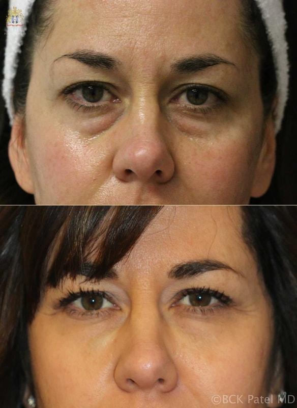
Correction of poorly placed filler causing a Tyndall effect and also lower eyelid bag exacerbation. A “Hammock Lift” with removal of residual filler, correction of the fullness, repositioning of the lower eyelid skin and correction of the zygomatic groove with nano fat grafts was carried out
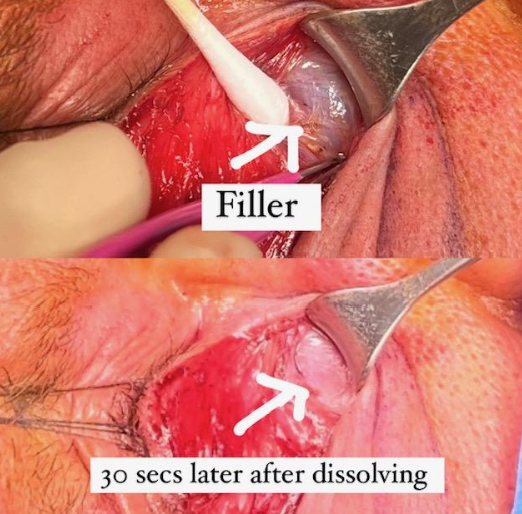
An example of an encapsulated area of filler which was dissolved directly suring surgery and the residual filler was surgically removed
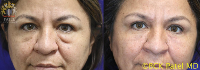
A long-lastig filler (hydroxyapatite) had been used in this patient resulting in lower eyelid firm fullness which was asymmetric. These fillers cannot be dissolved and require surgical removal. A Hammock lift with removal of the filler, repositioning of the lower eyelid skin laxity, repositioning of the cheeks and filling of the malar groove and the cheek volume loss with nano fat grafts was carried out.
Choosing the Right Injector Matters
A common factor across each of these complications is the injector. Working with a skilled injector can minimize the risk of each potential complication. In contrast, an inexperienced injector may increase the risk.
With the under-eye area, patients need a clinician with specialized experience. At his plastic surgery center in Salt Lake City, Dr. Patel provides comprehensive care as an oculoplastic surgeon. He understands that the under-eye area is one of the more technically demanding areas for injectable treatment.
References:Navigating Lower Eyelid Blepharoplasty in Patients with Under-Eye Filler: Challenges, Complications, and Recommendations. Yao A, Shah-Desai S, Malhotra R, Patel B.C.K. Plast Reconstr Surg. 2025 Dec 8. doi: 10.1097/PRS.0000000000012686.
Hyaluronic acid gel (Restylane) filler for facial rhytids: lessons learned from American Society of Ophthalmic Plastic and Reconstructive Surgery member treatment of 286 patients. McCracken MS, Khan JA, Wulc AE, Holds JB, Fante RG, Migliori ME, Ebroon DA, Amato MM, Silkiss RZ, Patel BCK. Ophthalmic Plast Reconstr Surg. 2006 May-Jun;22(3):188-91. doi: 10.1097/01.iop.0000217562.64529.ff.