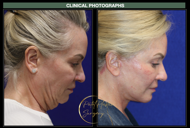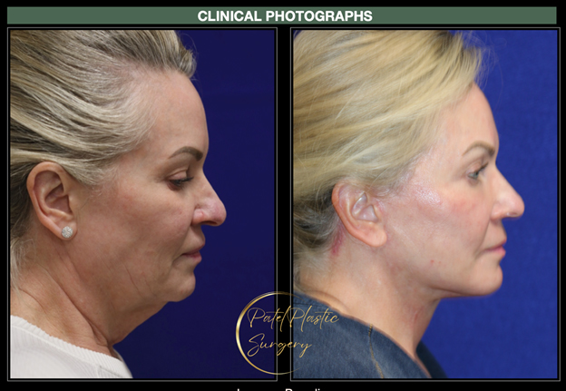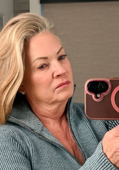
The appearance of the face and even the jawline and neck is different day to day. Overnight, fluid leakage into the tissues causes puffy upper and lower eyelids and even puffy jowls. Similarly, diet (e.g., a salty meal, wine), tiredness (lack of sleep), stress, and other factors can also affect how we look. Since we are generally examining patients during the daytime when patients are fresh, we are not seeing them at their worst. To that end, we encourage our patients to take photos of their face (and jawline and neck) when they have a “bad” day.
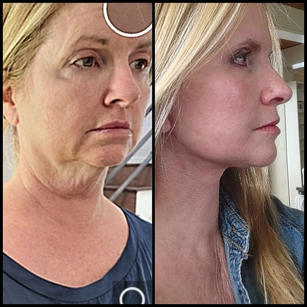
Also, everyone has looked at photographs taken by others when we erase the photograph because “I don’t like how I look.” These are also the photographs that we like to review. By assessing your face with our own measurements and photographs, but also by looking at your face, jawline and neck on “bad” days, we can plan your surgery more accurately. You will still have fluid leakage and good and bad days. But with a proper review of the “bad” photographs, we can design the deep plane facelift and neck lift to make these changes less extreme.