
Marking the Safe Zone when performing a Temporal Artery Biopsy
The anterior temporal hairline is lateral to the frontalis muscle; therefore, dissecting superior or posterior to the anterior hairline during a temporal artery biopsy is unlikely to injure the temporal branch of the facial nerve. However, because there are no muscles of facial expression in the temple region, only the thin superficial temporal fascia, the temporal branch of the facial nerve is particularly susceptible to injury over the zygoma and within the temple region.
Some authors define the danger zone for the temporal branch of the facial nerve by outlining a region bounded inferiorly by a line from the earlobe to the lateral eyebrow and superiorly by a line from the intertragal notch or earlobe to the lateral edge of the highest forehead crease. Other authors define up to 1.5 cm posterior to the lateral orbital rim and up to 1 cm anterior to the attachment of the helix along the level of the zygoma as safe zones to avoid nerve injury.[ See the video for an example of how to mark the safe zone preoperatively, based on cadaver studies by Shin et al.
Biopsy Site
Some authors recommend obtaining the biopsy specimen from the trunk of the superficial temporal artery, proximal to the division into frontal and parietal branches. Others feel that this technique sacrifices an unacceptably large arterial zone. Branches of the superficial temporal artery are useful for perfusing local and regional craniofacial reconstruction flaps and transferring free flaps to repair large facial defects. For this reason, it may be more prudent to limit the extent of superficial temporal artery sacrifice to an anterior branch. Other surgeons have suggested performing the biopsy on the parietal branch of the superficial temporal artery to eliminate the chance of injury to the temporal branch of the facial nerve. The parietal branch of the artery travels subperiosteally after separating from the main trunk of the superficial temporal artery. The sensitivity and specificity of parietal branch specimens are not known. However, the parietal branch of the superficial temporal artery provides collateral circulation to the brain, which may warrant preservation.
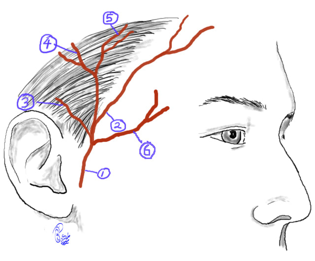
Superficial Temporal Artery: 1. Superficial Temporal Artery 2. Anterior (frontal) branch 3. Anterior auricular branch 4 & 5. Parietal branches 6. Middle temporal. Contributed by Prof. Bhupendra C. K. Patel MD, FRCS
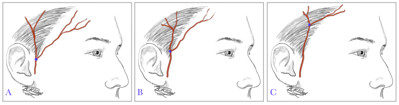
Superficial Temporal Artery: points of bifurcation. A. Low type just above the tragus (7%) B. Intermediate type (20%) C. High type (72%). Contributed by Prof. Bhupendra C. K. Patel MD, FRCS
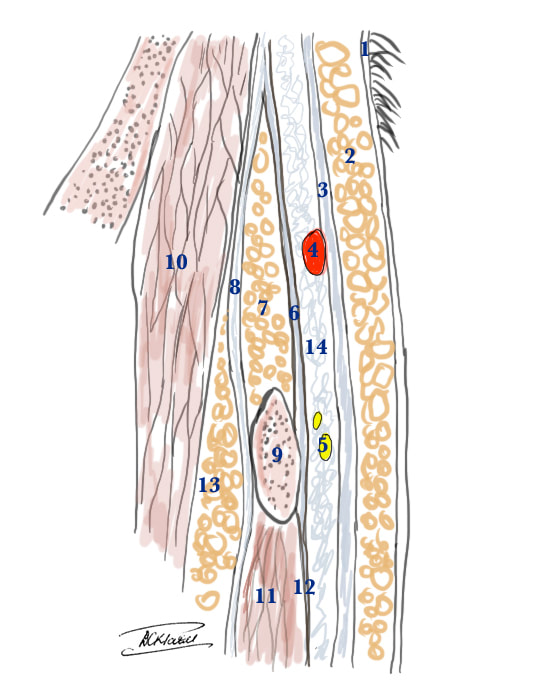
Superficial Temporal Artery and the Temporal branch of the Facial Nerve: anatomical cross section to show the relative layers above the zygomatic arch 1. Skin 2. Subcutaneous fat 3. Superficial temporal fascia (also called temporoparietal fascia) 4. Temporal artery within the superficial temporal fascia 5. Temporal branch of the facial nerve is just a little deeper than the artery below the superficial temporal fascia 6. Superficial layer of deep temporal fascia 7. Superficial temporal fat pad 8. Deep layer of deep temporal fascia 9. Zygomatic arch 10. Temporalis muscle 11. Masseter muscle 12. Masceteric fascia. Contributed by Prof. Bhupendra C. K. Patel MD, FRCS